atrial septal defect qp/qs
|
CHD Clinical Practice Algorithm: PFO/Atrial Septal Defect < 5
CHD Clinical Practice Algorithm: Atrial Septal Defect Post-Intervention10 Inclusion Criteria: • After surgical or cath based ASD closure Cath Lab Closure Or Surgical Surgical Cath Standard Surgical Follow up: Post-op ECG echo and CXR done prior to discharge Clinic follow-up within 2 weeks to 3 months with echo +/- ECG and CXR |
What should Qp and QS be in ASD?
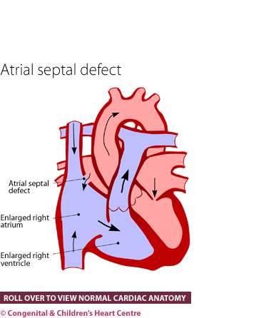
In the case of an isolated ASD (i.e. no other shunts or regurgitant valve disease), Qp should equal the RV stroke volume, and Qs should equal the LV stroke volume. Qp/Qs will be >1 in most cases because left atrial pressure typically exceeds right atrial pressure.
What is the Qp/Qs of a right atrial shunt?
Qp/Qs will be >1 in most cases because left atrial pressure typically exceeds right atrial pressure. However, in patients with pulmonary hypertension and right ventricular hypertrophy (i.e. Eisenmenger physiology), it is possible for the shunt to reverse and be predominantly right-to-left, resulting in a Qp/Qs < 1.0.
How do you know if you have a secundum atrial septal defect?
Diagnosed via echo with a secundum atrial septal defect Simple ASD w/o comorbiditiesExclusion Criteria: Pregnancy Dilated right sided structures History of stroke or prothrombotic state Clinic Visit ECG Echocardiogram1 After surgical or cath based ASD closure Post-op ECG, echo and CXR done prior to discharge
Why Cardiac MRI Is Beneficial
Cardiac MRI is well suited for the examination of an ASD because it can accurately quantify the shunt, and its effect on cardiac structure and function. It can directly visualize blood flowing across the ASD. It is totally non-invasive and does not require contrast. cardiacmri.com
MRI Technique
Short and long axis SSFP images are obtained to quantify the end-diastolic and end-systolic volumes of both ventricles, and to determine global systolic function (LV and RV EF). Phase contrast images are acquired to quantify blood flow in the main pulmonary artery (Qp) and the ascending aorta (Qs) to determine the shunt ratio (Qp/Qs). For the most
IV. Analysis
The best way to quantify the shunt ratio (Qp/Qs) is to divide the blood flow in the main pulmonary artery (Qp) by the blood flow in the ascending aorta (Qs). In the case of an isolated ASD (i.e. no other shunts or regurgitant valve disease), Qp should equal the RV stroke volume, and Qs should equal the LV stroke volume. Qp/Qs will be >1 in most cas
v. Which Imaging Findings Affect Treatment?
The severity of ASD can be quantified in terms of Qp/Qs and defect size. Percutaneous closure and surgery are generally reserved for patients with Qp/Qs > 1.5, unless they are undergoing cardiac surgery for other reasons. cardiacmri.com
VI. Drawbacks of Existing Tests
Transthoracic echocardiography is most commonly used to assess for an ASD. Many patients also undergo transesophageal echocardiography for a better anatomic assessment. MRI is less invasive and is superior for quantifying the size of the shunt (Qp/Qs), as well as its affect on RV size and function. MRI is also good at identifying other possible ass
|
Pathophysiology of Congenital Heart Disease in the Adult
26 Feb 2008 If the left-to-right shunt equals the right-to-left shunt in magni- tude it is possible to have a Qp/Qs of exactly 1:1. ... atrial septal defect ... |
|
Natural history of medium-sized atrial septal defect in pediatric cases
22 Jun 2012 Background: The indication for surgical repair of atrial septal defect (ASD) is pulmonary to systemic blood flow ratio (Qp/Qs) > 2.0 ... |
|
Cardiac MRI: Part 1 Cardiovascular Shunts
27 Dec 2010 aortic (Qs) and pulmonary (Qp) flows respec- tively |
|
Cardiac MRI: Part 1 Cardiovascular Shunts
27 Dec 2010 aortic (Qs) and pulmonary (Qp) flows respec- tively |
|
Transatrial Septal Velocity Measurement by Doppler
Catheterization Qp:Qs. t Radionuclide shunt scan. t Tricuspid flow method for Doppler Qp measurement. ASD = atrial septal defect; BSA = body surface |
|
Atrial Septal Defect in Patients over the Age of Forty Years
tests Qp/Qs was determined by oximetry. Oxy- gen contents were determined in PA |
|
Noninvasive evaluation of the ratio of pulmonary to systemic flow in
monary arterial flow inpatients with atrial septal defect. (ASD) and/or with ASD Qp/Qs ranged from 0.51 to 4.6. Seven patients had pulmonary hypertension ... |
|
Doppler color-flow imaging assessment of shunt size in atrial septal
symbol sinus venosus defect; E |
|
Accuracy of doppler echocardiography in quantification of left to right
septal defect the Qp/Qs ratio by oximetry was compared There wa a good correlation between Doppler and oximetric. Qp/Qs mtios in patients with atrial septal ... |
|
What radiologists need to know about the pulmonary---systemic flow
We describe the measurement of Qp/Qs in simple intracardiac shunts (septal defects). Predictors of spontaneous closure of isolated secundum atrial septal ... |
|
Pathophysiology of Congenital Heart Disease in the Adult
26 févr. 2008 If the left-to-right shunt equals the right-to-left shunt in magni- tude it is possible to have a Qp/Qs of exactly 1:1. Atrial Septal Defect. |
|
Natural history of medium-sized atrial septal defect in pediatric cases
22 juin 2012 Background: The indication for surgical repair of atrial septal defect (ASD) is pulmonary to systemic blood flow ratio (Qp/Qs) > 2.0 ... |
|
Simple Atrial Septal Defects What you need to look for..
_gina_-_simple_atria.pdf |
|
Doppler color-flow imaging assessment of shunt size in atrial septal
left-to-right (Qp/Qs) shunts. In a study of 39 ASD patients with a small (<2: 1) left-to-right shunt who were followed for 5-21 years (mean |
|
2019-03- XAPLANTERIS -Septal defects - 2
Atrial septal defects Septal defects result in L to R shunts ... Qp:Qs = 23$(/ %'-4)'*/01 |
|
Noninvasive evaluation of the ratio of pulmonary to systemic flow in
duplex Doppler echocardiography in 22 patients with atrial septal defects (ASDs). Right and left mining Qp/Qs in patients with ASD by comparing. |
|
Effect of atrial septal defect repair on left ventricular geometry and
30 août 2016 TABLE 1. Clinical and Hemodynamic Features of the Study Group. Age. Associated. PAP. Pt. (years). Sex. Type ASD defect. Qp/Qs. |
|
Transcatheter Atrial Septal Defect Device Closure - A 2.5 Years
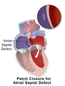
Transcatheter device closure is advised for all symptomatic patients and also for asymptomatic patients with a Qp: Qs ratio of at least 2:1 or those with right |
|
Non-invasive dye dilution method for measuring an atrial septal
5 oct. 2020 quantify shunt size related to atrial septal defects (ASD). ... ASD. Mean pulmonary blood flow/systemic blood flow. (Qp/Qs) was measured ... |
|
Echocardiography in the Assessment of Atrial Septal Defects
She was diagnosed with a secundum atrial septal defect. (Please see companion DVD for corresponding video.) Fig. 5. Quantification of shunt flow (Qp:Qs) |
|
ATRIAL SEPTAL DEFECT - NCBI
Patent foramen ovale and atrial (or auricular) septal defect (A S D ), though both characterized by an aperture in the atrial septum, are embryologically and |
|
Atrial septal defects - CORE
9 avr 2014 · Secundum atrial septal defect is a defect within the fossa ovalis usually due to one or several defects within septum primum (figure 2B) Septum |
|
Atrial septal defect - Great Ormond Street Hospital
The atrial septum is the wall of tissue and muscle between the upper two chambers (atria) of the heart An atrial septal defect (ASD) is a hole in this wall |
|
Understanding your childs heart Atrial septal defect - British Heart
What is atrial septal defect? An atrial septal defect (known as ASD) is a hole between the two upper chambers (atriums) of the heart |
|
ATRIAL SEPTAL DEFECT - BMJ Heart
In atrial septal defect the mean pressure in the right ventricle and pulmonary artery may be normal or little above normal in the presence of grade III splitting It is |
|
Atrial Septal Defect - American Heart Association
An ASD is an opening or hole (defect) in the wall (septum) between the heart's two upper chambers (atria) What causes it? Every child is born with an opening |
|
Atrial Septal Defect
The secundum atrial septal defect (ASD) is the second most common form of congenital heart disease, representing at least 10 of all congen- ital cardiac |


![Atrial Septal Defect (ASD) - [PDF Document] Atrial Septal Defect (ASD) - [PDF Document]](https://f1.media.brightcove.com/8/3850378299001/3850378299001_5536892101001_5536883677001-vs.jpg?pubId\u003d3850378299001\u0026videoId\u003d5536883677001)


![PDF] Congenital secundum atrial septal defect and membranous PDF] Congenital secundum atrial septal defect and membranous](https://imgv2-2-f.scribdassets.com/img/document/325305910/298x396/f914aa37f4/1474865659?v\u003d1)


