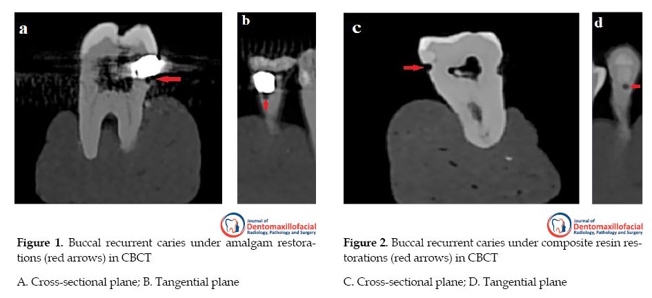
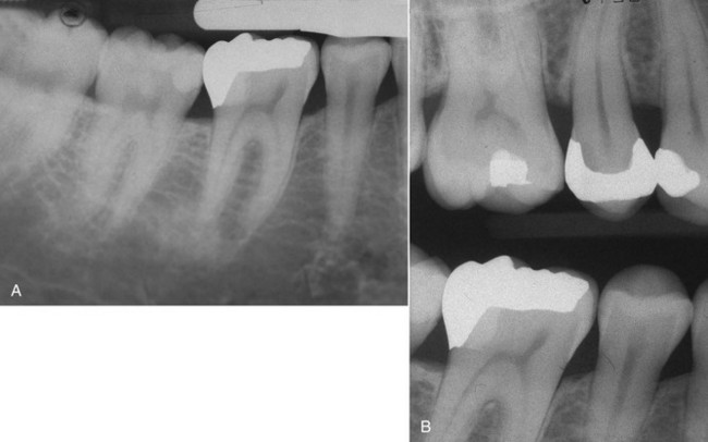
amalgam restoration radiograph
What does amalgam look like on xray?
Tooth enamel and metallic restorations (amalgams, crowns, etc.) are very dense, and deflect X-‐rays preventing them from reaching the film.
Tooth enamel and amalgams look white (radiopaque).Amalgam (most radiopaque)SS crown.Gutta percha.Acyclic resin(least radiopaque)
Is amalgam restoration radiopaque or radiolucent?
In the past, standard dental restorative materials were metals such as amalgam, gold or non-precious alloys with high atomic numbers.
These metals – i.e. titanium, copper, strontium, ytterbium, silver, gold, bromine, barium, and strontium – are radiopaque and easily block out x-rays.
How will an amalgam filling appear on a radiographic image?
Denser areas– such as amalgam fillings and other restorations – will block most of the photons and appear white, while teeth and tissue will show in shades of gray.
|
Progression of Radiopacities and Radiolucencies under Amalgam
Dec 14 1995 Amalgam restoration. Bite-wing radiographs. Radiopaque and radiolucent dentine. Secondary caries diagnosis. Abstract. Radiolucent and radiopaque ... |
|
Progression of Radiopacities and Radiolucencies under Amalgam
Radiolucencies under Amalgam. Restorations on Bite-Wing. Radiographs. Key Words. Amalgam restoration. Bite-wing radiographs. Radiopaque and radiolucent dentine. |
|
Chapter 3 - Radiographic Technique
Apr 3 2014 Tooth enamel and metallic restorations (amalgams |
|
Clinical and Radiographic Assessment of Class II Esthetic
Radiograph of mesio-occlusal Vitremer restoration In contrast glass ionomer and amalgam restorations presented significantly less radiographic defects at the ... |
|
A Comparison of the Accuracy of Digital and Conventional
Dec 1 2010 Validity of radiographs for diagnosis of secondary caries in teeth with class II amalgam restorations in vitro. Caries. Res. 1997; 31(1):24-9. |
|
A deep learning approach to dental restoration classification from
2020) with amalgam composite resin |
|
Detection of marginal defects of composite restorations with
marginal defects of composite resin restorations based on radiographs was only slightly affected by the radiographic system being used. The diagnosis of |
|
Overhanging Amalgam Restorations by Undergraduate Students
Key Words: Overhanging amalgam margins. Dental amalgam. Class-II restoration. Bitewing radiograph. 1 Department of Operative Dentistry Dow Dental College |
|
Radiographic Assessment of Primary Molar Pulpotomies Restored
56 The success of amalgam restorations to restore pulpotomized primary molars was previously re- ported |
|
Radiographic Success of Ferric Sulfate and Formocresol
amalgam restoration. Radiographic findings. Using the radiographic criteria outlined in the Methods section 15 of the 35 teeth (43%) treated with ferric sul |
|
Progression of Radiopacities and Radiolucencies under Amalgam
14 déc. 1995 Amalgam restoration. Bite-wing radiographs. Radiopaque and radiolucent dentine. Secondary caries diagnosis. Abstract. |
|
Cross-sectional radiographic survey of amalgam and resin-based
Cross-sectional radiographic survey of amalgam and resin-based composite posterior restorations. Liran Levin DMD1/Marius Coval |
|
Overhanging Amalgam Restorations by Undergraduate Students
Dental amalgam. Class-II restoration. Bitewing radiograph. 1 Department of Operative Dentistry Dow Dental College |
|
Fuks 5431
composite resin restorations presented radiographic defects that might require replacement at a later date. In contrast glass ionomer and amalgam |
|
Examiner Agreement in the Replacement Decision of Class I
15 mai 2004 replacement decision of Class I amalgam restorations based on clinical and radiographic ... amalgam restoration using a dental X-ray unit. |
|
Assessment of Different Techniques to Detect Recurrent Carious
Background: This in-vitro study was to evaluated bitewing radiograph and tactile examination for detection secondary caries adjacent to amalgam restorations |
|
The Prevalence of Amalgam Overhang in Erbil City Population
showed that the prevalence of overhang dental restorations is very significant and alarmingly radiograph was taken to evaluate the amalgam restoration. |
|
Accuracy of various imaging methods for detecting misfit at the tooth
tooth-restoration interface: (1) bitewing radiographs both conventional and digital |
|
A deep learning approach to dental restoration classification from
the detection and differentiation of amalgam composite resin |
|
Chapter 3 - Radiographic Technique
3 avr. 2014 The goal in dental radiology is to use techniques that require the least amount of ... The lightest areas are amalgam restorations. |
|
Radiographic analysis of various restorations on teeth – An in vitro
Class 1 cavity preparation was done using a 245 bur The teeth were then randomly allocated into three groups of 10 each and restored with amalgam, composite, |
|
The radiographie investigation of the visibility of secondary caries
The teeth were restored whh amalgam The teeth were adapted in the actual lootli space of 15 volunteers with one mandibular premolar missing Radiographs |
|
Evaluation of the Radiopacity of Different Restorative Materials by
Keywords: composite resins; glass ionomer cements; radiographic image Interestingly, one of the first recommendations for those restorations was the use of a |
|
A radiographic and scanning electron microscopic study of
Clinical studies on the quality of Class II amalgam and resin composite restorations frequently report defective cervical margins A&: The aim of this study was to |
|
Overhanging Amalgam Restorations by Undergraduate - JCPSP
Key Words: Overhanging amalgam margins Dental amalgam Class-II restoration Bitewing radiograph 1 Department of Operative Dentistry, Dow Dental |
|
Clinical investigation of a radiopaque composite - GovInfo
commercially available radiolucent composite (Fig 1) Figure 2 shows four different radiographs of the same extracted tooth The restoration has been replaced |
![PDF] Clinical and radiographic assessment of Class II esthetic PDF] Clinical and radiographic assessment of Class II esthetic](https://j.kwikweb.co.za/jtm/photos/Figure%2015.jpg)


![PDF] Frequency of Iatrogenic Changes Caused from Overhang PDF] Frequency of Iatrogenic Changes Caused from Overhang](https://casereports.bmj.com/content/bmjcr/12/9/e230248/F1.large.jpg)